Cow Diseases and Symptoms: Complete Guide With Pictures & Treatment (2025)
- BeyondForest

- Nov 10, 2025
- 16 min read
Updated: Nov 28, 2025

1.)Most Common Cow Diseases and Their Symptoms
4.)Parasitic & Tick-Borne Diseases
6.)Cow Diseases and Symptoms Pictures
8.)Emergency First Aid for Sick Cattle
Mastitis |
Foot and Mouth Disease (FMD) |
East Coast Fever (ECF) |
Lumpy Skin Disease (LSD) |
Milk Fever |
Anthrax |
Bloat |
Pneumonia |
Brucellosis |
Trypanosomiasis (Nagana) |
Mastitis
Mastitis is one of the most common and costly dairy cow diseases, caused by bacterial infection of the udder tissue. It reduces milk production, affects milk quality, and can permanently damage the udder if not treated early.

Causes of Mastitis
Euthanasia is the practice of intentionally ending a life to eliminate pain and suffering.
Dirty milking environment | contaminated bedding, wet floors, muddy kraals. |
Poor milking hygiene | unclean hands, towels, or milking machines |
Udder injuries | cuts, bruises, or teat damage. |
Incomplete milking | leaving milk in the udder encourages bacterial growth. |
Cow immunity issues | stress, poor nutrition, or post-calving vulnerability. |

Visible (Clinical Mastitis):
Swollen, hot, or painful udder quarters
Watery, clotted, thick, or bloody milk
Reduced milk yield
Cow avoids milking due to pain
Fever, loss of appetite (in severe cases)
Hidden (Subclinical Mastitis):
There is no obvious signs, but Milk production drops, Milk becomes watery and Somatic cell count (SCC) rises (Subclinical mastitis is responsible for 70–80% of losses because farmers don’t notice it early.)

How to Treat Mastitis
Treatment should begin immediately to avoid permanent udder damage.
Intra-mammary antibiotics
Most common treatment (e.g., intramammary tubes).
Must follow withdrawal periods to keep milk safe.
Anti-inflammatory drugs
Reduce pain and swelling.
Frequent stripping
Helps remove bacteria and infected milk.
Vet diagnosis
Use California Mastitis Test (CMT) to identify which quarter is affected.
Clean cows’ bedding daily (dry, soft environment). |
Wash and sanitize teats before and after milking. |
Use separate clean towels for each cow. |
Maintain and disinfect milking machines regularly. |
Milk healthy cows first, infected cows last. |
Provide balanced nutrition to boost immunity. |
Dry cow therapy after lactation. |
Milk Fever
Milk Fever (also called Hypocalcemia) is a metabolic disease that occurs when a cow’s blood calcium levels drop suddenly, usually within 24–72 hours after calving. It is common in high-producing dairy cows and can be fatal if not treated quickly.

Image of a Cow with Milk Fever By Dairy Verse
Sudden high demand for calcium | during colostrum and early milk production |
Poor mineral balance | in the diet before calving |
Older cows | 3rd lactation and above are more at risk |
Low magnesium levels | which reduce the cow’s ability to absorb calcium |
Feeding too much calcium pre-calving | , making the body “lazy” at mobilizing calcium |

Symptoms of Milk Fever
Milk Fever progresses in three stages. Early recognition saves the cow.
Stage 1 (Mild)
Restlessness
Muscle tremors
Stiff walking
Loss of appetite
Stage 2 (Moderate) — Most Common
Cow sits in a “S-shaped” neck position
Cold ears and cold muzzle
Weak heartbeat
Cow cannot stand
Dry muzzle, constipation
Stage 3 (Severe/Advanced) — Emergency
Cow lies flat on her side
No muscle control
Low body temperature
Risk of bloat or death within hours
Treatment must begin immediately with
IV Calcium Borogluconate (Most Effective)
Given slowly into the vein by a trained person or veterinarian.
Cow usually stands within 30 minutes.
Subcutaneous Calcium
Given under the skin in multiple sites for slower absorption.
Oral Calcium Gel or Drench
For mild cases or after IV treatment to prevent relapse.
Warm the cow
Use blankets or warm water bottles.
Prevents hypothermia during recovery.
Low-calcium diet 3 weeks before calving(forces the body to learn how to mobilize calcium) |
Add magnesium to pre-calving diet |
Avoid feeding dairy meal too early before calving |
Provide oral calcium immediately after calving |
Balance rations with a nutritionist |
Ensure cows are at the correct body condition score (BCS 3–3.5) |
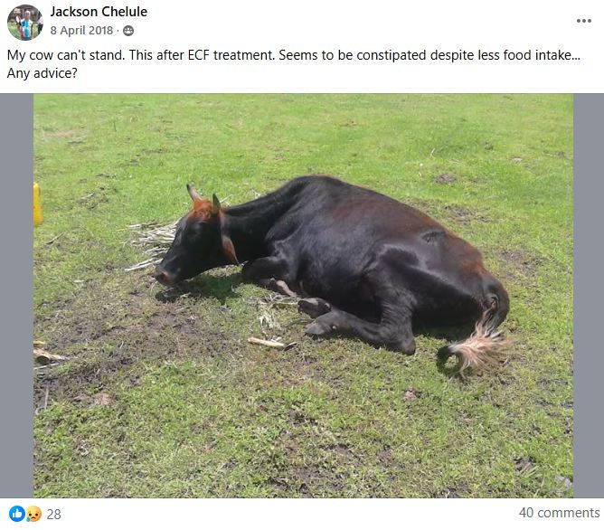
East Coast Fever
East Coast Fever (ECF) is one of the deadliest cattle diseases in East Africa, caused by a protozoan parasite (Theileria parva) transmitted through the brown ear tick. It kills thousands of cattle every year, especially calves and exotic breeds, and can destroy entire herds if not controlled early.

ECF spreads when infected brown ear ticks feed on cattle.
Tick bites on ears, tail switch, udder, dewlap & legs |
Poor tick control on farms |
Mixing animals from different farms without quarantine |
Unvaccinated calves (highest risk) |
Frequent grazing in tick-infested grasslands |
The disease is common in warm, humid, high-tall-grass areas where ticks thrive.

Symptoms appear 10–18 days after the tick bite.
Early Symptoms
High fever (up to 41°C) |
Swollen lymph nodes (especially parotid glands behind the ear) |
Loss of appetite |
Lethargy / weakness |
Progressive Symptoms
Coughing |
Difficulty breathing |
Nasal discharge |
Watery eyes |
Weight loss |
Critical/Advanced Stage
Severe respiratory distress |
Cow collapses and cannot stand |
“Pneumonia-like” signs as lungs fill with fluid |
Death within 3–7 days if untreated |
Treatment must be done immediately once ECF is suspected.
1. Buparvaquone (Butalex or Bupaject)
Most effective ECF drug
Must be given by a vet
Works best in the early stage
2. Oxytetracycline
Used for mild cases or supportive treatment
3. Supportive Therapy
Anti-inflammatories
Multivitamins
Fluid therapy
Good feeding to boost immunity
Note ECF treatment is expensive because the drug itself is costly and late-stage survival is low.

Good prevention is cheaper than treatment.
Tick Control
Weekly or twice-weekly spraying with acaricides |
Pour-on tick control |
Hand-picking and inspecting calves regularly |
Clearing tall grass around sheds |
ECF Vaccine (Infection & Treatment Method – ITM)
Long-term protection |
Given once in a lifetime |
Best for calves 3–9 months |
Must be administered by trained vets |
Farm Management
Quarantine new cattle for 21 days |
Keep exotic breeds in well-managed tick-free systems |
Provide balanced nutrition to boost immunity |
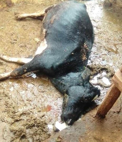
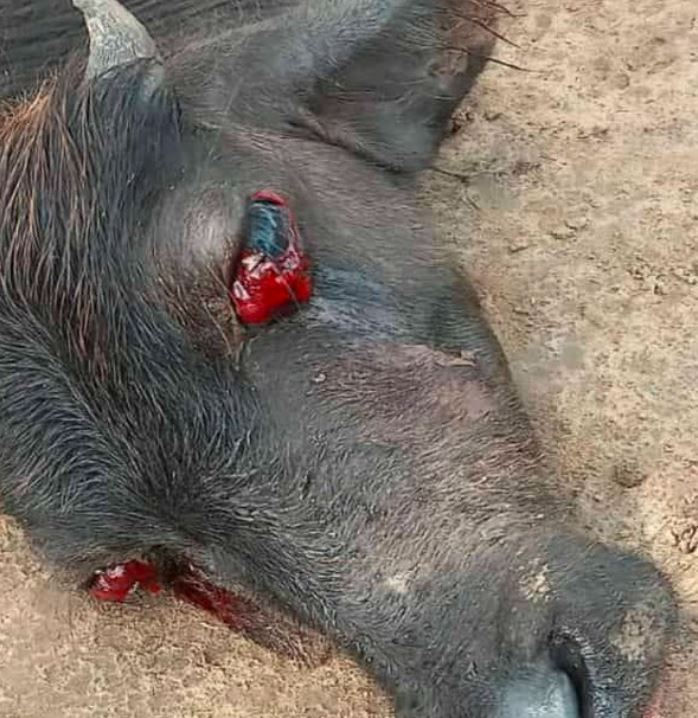
Do NOT open the carcass. Opening it exposes the bacteria to oxygen → the bacteria forms spores → the land becomes contaminated for decades.
Anthrax is one of the most dangerous bacterial diseases in livestock, caused by Bacillus anthracis. It leads to sudden death, spreads rapidly, and can infect humans. Because it is a notifiable disease, any suspected case must be reported to veterinary authorities immediately.
If you see a cow dead with blood leaking from openings → Anthrax is the first suspect.
Anthrax spreads from spores found in contaminated soil, water, feed, and carcasses. The spores can survive for 30–50 years, especially in areas with past outbreaks.
Grazing on contaminated pasture |
Drinking water from infected streams or ponds |
Opening or cutting carcasses of animals that died suddenly |
Poor carcass disposal |
Rainy seasons that bring spores to the surface |
Anthrax is unique because cows often die suddenly with very few signs. However, when symptoms appear, they include:

Early Symptoms (1–2 hours before death)
Sudden high fever
Difficulty breathing
Trembling or staggering
Swelling of the neck, chest, or belly
Classic Signs After Death

Blood oozing from the nose, mouth, and anus
Blood does NOT clot (remains dark and watery)
Rapid bloating of the carcass
No rigor mortis (body stays soft)
Treatment
High-dose Penicillin |
Oxytetracycline |
Strict isolation of sick animals |
Supportive care |
Best Way to Protect Your Herd against Anthrax
Annual Vaccination
Use the Stern Anthrax vaccine |
Given once per year |
Cheapest and most effective prevention |
Don’t vaccinate sick animals or those under antibiotic treatment |
Proper Carcass Disposal
Burn or bury 2 meters deep |
Apply lime on disposal site |
Farm Biosecurity
Keep livestock away from old burial pits |
Avoid grazing in known anthrax-prone areas during rainy seasons |
Quarantine new animals for 14 days |

Trypanosomiasis, also known as Nagana, is a deadly parasitic disease transmitted mainly by tsetse flies. It affects cattle, camels, goats, and wildlife, causing severe weight loss, anemia, low milk yield, and eventually death if untreated. It is common in tsetse-infested regions across Africa.
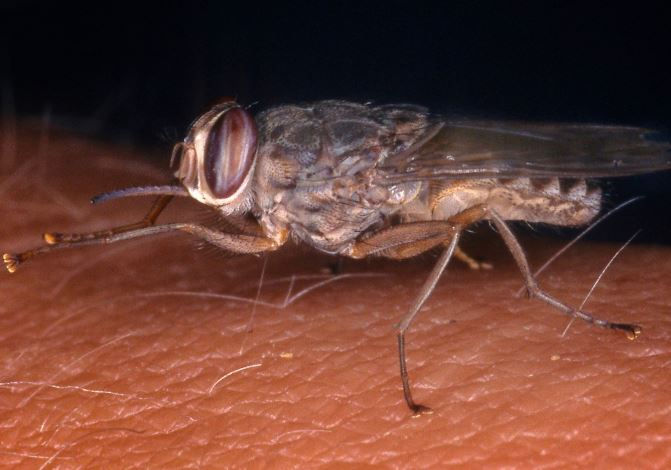
The disease is caused by parasitic protozoa
Trypanosoma congolense |
Trypanosoma vivax |
Trypanosoma brucei |
Transmission occurs through Tsetse flies (main vector), Biting flies like tabanids and stable flies and sharing needles during treatment. The parasites multiply in the blood, lymph, and tissues, damaging major organs.
Symptoms of Trypanosomiasis in Cattle
Symptoms develop gradually over weeks.
Early Symptoms
Fever
Loss of appetite
Depression / weakness
Enlarged lymph nodes (especially behind the jaw)
Progressive Symptoms
Severe weight loss (“eaten from inside”)
Drop in milk yield
Rough, dull hair coat
Pale or yellowish mucous membranes
Labored breathing
Edema under the jaw (“bottle jaw”)
Final Stage
Emaciation
Recumbency (cow cannot stand)
Organ failure
Death if untreated
How to Treat Trypanosomiasis
Treatment must be fast and strategic.
Diminazene Aceturate
(Commercial names: Berenil®, Veriben®)
Most commonly used drug
Effective for T. congolense and T. vivax
Isometamidium Chloride
(Commercial names: Samorin®, Trypamidium®)
Used for treatment and prevention
Offers 2–3 months protection in high-risk areas
Homidium Salts
(Homidium Bromide or Homidium Chloride)
Used in some regions for early/mild infections
Supportive Therapy
Multivitamins
Iron supplements
Deworming
Good nutrition
Tsetse Fly Control
Spraying cattle with pyrethroid-based acaricides
Pour-ons (e.g., Cypermethrin, Deltamethrin)
Insecticide-impregnated traps
Bush clearing in grazing areas
Keeping cattle away from tsetse zones in early morning & late afternoon (peak bite times)
Prophylactic Treatment
Isometamidium given every 2–3 months in high-risk areas
Good Farm Management
Quarantine new animals
Avoid sharing needles
Regular deworming & nutrition to boost immunity
FMD |
Lumpy Skin Disease |
Rabies |
Rift Valley Fever |
Bovine Viral Diarrhea (BVD) |
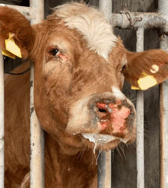
Foot and Mouth Disease

Foot and Mouth Disease (FMD) is one of the most contagious viral diseases affecting cattle, sheep, goats, and pigs. It spreads extremely fast, causes huge economic losses, and can wipe out livestock in entire regions if not controlled. Although adult cows rarely die, the disease causes severe productivity losses and high calf mortality.
Causes of Foot and Mouth Disease
FMD is caused by the FMD virus (FMDV), which has 7 serotypes. Infection spreads through
Direct contact with infected animals |
Contaminated feed, water, or equipment |
Milkers, workers, vehicles, shoes |
Airborne transmission (up to 10 km in some climates) |
Raw milk and meat from infected animals |
Foot and Mouth Disease virus survives in soil, manure, milk, and the environment for long periods.

Symptoms of FMD in Cattle
Symptoms appear 2–14 days after exposure.
Mouth Lesions
Drooling or excessive saliva (“ropey saliva”)
Blisters on Tongue, Gums ,Lips ,Inside cheeks
Mouth pain → difficulty eating → sudden drop in appetite
Foot Lesions
Blisters around Hoof coronet ,Between the toes, Heel bulbs
Lameness (cow refuses to walk)
Swollen or painful feet
Milk & Production Changes
Sudden drop in milk yield (up to 80%)
Fever
Depression / weakness
In Calves
Heart damage (“tiger heart lesions”)
Sudden death even without mouth or foot blisters

No direct cure because FMD is viral. Treatment is supportive:
Supportive Care
Anti-inflammatories to reduce pain
Antibiotics to prevent secondary infections
Fluids and energy supplements for calves
Soft feed and plenty of water
Mouth & Foot Care
Clean blisters with antiseptic
Apply gentian violet, iodine, or aloe vera
Keep animals on soft ground to reduce pain
Isolation
Sick animals must be isolated immediately
Healthy animals should not share water troughs or feeding points

Prevention & Control of FMD
Vaccination (Most Important)
Regular vaccination depending on region |
Boosters required based on the circulating strains |
Essential for dairy and beef herds |
Movement Control
Quarantine new animals for 14–21 days |
Limit livestock movement during outbreaks |
Disinfect vehicles, shoes, and tools |
Farm Biosecurity
Footbaths at entrance |
Clean milking equipment |
Separate sick and healthy animals |
Restrict visitors to the farm |
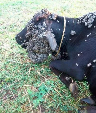
Lumpy Skin Disease

Image of a Cow with LMD by ThisFarmer Carson
Lumpy Skin Disease (LSD) is a highly infectious viral disease caused by the Capripoxvirus. It affects cattle of all ages and breeds and is spread mainly by biting insects. The disease causes painful skin nodules, fever, reduced milk yield, and severe economic loss in dairy and beef herds.
Causes of Lumpy Skin Disease
LSD spreads through
Biting insects – mosquitoes, flies, ticks |
Direct contact between cattle |
Contaminated tools, needles, halters, ropes |
Semen from infected bulls |
Movement of infected animals |
The virus survives long in scabs, skin, and the environment.

Image of a Calf infected by LSD by Demi Farms
Symptoms appear 6–14 days after infection.
Skin Nodules (Main Sign)
Hard, round lumps 1–7 cm on Neck , Back ,Legs ,Udder & teats ,Face & muzzle. Nodules may become deep wounds (sit-fasts)
General Symptoms
High fever
Loss of appetite
Swollen lymph nodes
Lethargy / weakness
Runny nose and eyes
Effects on Milk & Reproduction
Sharp drop in milk production
Mastitis due to teat lesions
Infertility in bulls
Abortions in pregnant cows
Severe Cases
Painful walking (lameness)
Deep ulcers with secondary infections
Maggot infestation if untreated
There is no direct cure because LSD is viral. Treatment focuses on:
Supportive Care
Pain and fever reducers (NSAIDs)
Antibiotics to prevent secondary bacterial infections
Good feeding, water, and shelter
Wound Management
Clean wounds with iodine or chlorhexidine
Apply wound sprays (aluminium spray, oxytetracycline spray)
Keep flies away to prevent maggots
Isolation
Isolate sick animals immediately
Disinfect all shared equipment

Vaccination (Most Important)
Use the LSD vaccine (e.g., Neethling strain vaccine) |
Annual vaccination recommended |
Boosters may be needed in outbreak areas |
Vector Control
Regular spraying with pyrethroids |
Use pour-ons (cypermethrin, deltamethrin) |
Clear bushy areas and stagnant water |
Use fly traps |
Farm Management
Quarantine new animals for 14–21 days |
Do not share needles |
Maintain hygiene in cattle sheds |
Rabies
Rabies is a fatal viral disease that affects the brain and nervous system of all warm-blooded animals, including cattle, goats, sheep, dogs, and humans. Once symptoms appear, rabies has a 100% death rate. Because it is zoonotic, humans can also get infected.

Causes of Rabies in Cattle
Rabies is caused by the Rabies lyssavirus and spreads through:
Bites from infected dogs (most common in East Africa)
Bites from wild animals such as:
Jackals
Foxes
Bats
Mongoose
Saliva entering wounds, scratches, or mucous membranes
Licking of open wounds by infected animals
Once the virus reaches the brain, symptoms begin — and death follows rapidly.
Symptoms of Rabies in Cattle
Symptoms appear 2 weeks to several months after a bite, depending on where the cow was bitten.
Rabies occurs in two forms:
Furious Rabies (Aggressive Form)
The cow becomes abnormally excitable
Sudden aggression |
Charging at people or animals |
Excessive bellowing |
Restlessness |
Attempting to bite or attack |
Increased sensitivity to light and sound |
Foaming at the mouth |
This form is highly dangerous to humans.

Dumb/Paralytic Rabies (Most Common in Cattle)
Drooping head |
Difficulty swallowing |
Paralysis of the hind legs |
Cow stands with legs spread wide |
Loss of coordination |
Depression or acting “drunk” |
Progressive paralysis → coma → death |
This is the form most farmers see. Most cows die within 2–5 days after symptoms begin.
How to Treat Rabies in Cattle
There is NO cure. Once symptoms appear, treatment is not effective.The animal will die. What to do instead
Isolate the cow immediately |
Do NOT let anyone touch saliva or mucous |
Call a vet or livestock officer |
Notify local public health teams |
If a person has been licked, scratched, or bitten by the cow → immediate hospital visit for post-exposure vaccine |
Never slaughter or eat a cow suspected of rabies.
Prevention and Control of Rabies
Vaccination
Rabies vaccine is the main protection
Given annually in high-risk areas
Very important for:
Dairy cows
Bulls
Calves
Animals near wildlife zones
Farms with many dogs
Control Dogs & Wildlife
Vaccinate all farm dogs yearly
Reduce stray dog interactions
Secure cattle at night
Avoid grazing near wildlife corridors
After a Bite
If a cow is bitten by a suspected rabid animal:
Wash the bite wound with lots of water + soap
Call a vet immediately
Give post-exposure rabies vaccine (yes, cattle can receive it)
Monitor for 3–6 months
Early vaccination after a bite can save the cow before symptoms begin.
Rift Valley Fever
Rift Valley Fever (RVF) is a highly infectious viral disease that affects cattle, sheep, goats, camels, and humans.
It spreads rapidly during heavy rains and flooding when mosquito populations explode. RVF is zoonotic humans can get infected making it a serious public health concern.
Causes of Rift Valley Fever
RVF is caused by the Rift Valley Fever virus (RVFV) and spreads through
Mosquito bites (main transmission route) |
Contact with blood, tissues, or aborted fetuses of infected animals |
Handling raw meat, placentas, or assisting births |
Contaminated water and mud after flooding |
Slaughtering or skinning sick animals |
The virus survives in mosquito eggs for years and emerges after heavy rains.
Symptoms of RVF in Cattle
Symptoms vary by age and severity.
Adult Cattle
High fever
Sudden drop in milk production
Nasal discharge
Weakness and lethargy
Abortion storms (pregnant cows abort 80–100%)
Diarrhea (sometimes bloody)
Jaundice (yellow gums/eyes)
Calves (Severe Form)
Very high fever
Rapid breathing
Weakness
Sudden death within 24–48 hours
Mortality rate can reach 70–90%
How to Treat Rift Valley Fever
There is NO direct cure, as RVF is a viral disease.
Treatment focuses on supportive care
Anti-inflammatories for fever and pain |
Fluid support for dehydration |
Antibiotics to prevent secondary bacterial infections |
Clean environment to reduce stress |
Provide high-energy feeds |
Isolation is important because the blood and tissues of infected animals are contagious.

Prevention and Control of Rift Valley Fever
Vaccination
The most effective protection
Use the RVF livestock vaccine (live or inactivated)
Vaccinate animals before rainy seasons
Do NOT vaccinate pregnant cows with live vaccine

Mosquito Control
Spray cattle with insecticides (pyrethroids)
Remove stagnant water around sheds
Use pour-ons and insect repellents
Trim tall grass near watering points
Farm Biosecurity
Avoid slaughtering sick animals
Wear gloves when handling births or abortions
Burn or bury aborted fetuses and placenta
Quarantine new animals
Movement Control
RVF outbreaks lead to government bans on:
Livestock movement
Slaughter
Milk sale
Market activities
Bovine Viral Diarrhea
Bovine Viral Diarrhea (BVD) is a highly contagious viral disease that affects cattle of all ages. It weakens the immune system, causes reproductive losses, diarrhea, respiratory disease, and can lead to death in calves.
BVD is a major economic disease in dairy and beef herds.
Causes of BVD
BVD is caused by the Bovine Viral Diarrhea Virus (BVDV), which spreads through
Direct contact with infected cattle |
PI animals (Persistently Infected calves – major spreaders) |
Contaminated feed, water, equipment |
Semen from infected bulls |
Fetal infection during pregnancy |
Shared needles and instruments |
Persistently infected animals shed the virus every second of their life.
Symptoms of BVD in Cattle
Symptoms vary depending on age, immune status, and virus strain.
Acute BVD (Most Common Form)
Fever
Watery diarrhea
Nasal discharge
Coughing
Loss of appetite
Depression
Dehydration
Drop in milk production
Mucosal Disease (Deadly Form)
Occurs when a PI animal gets superinfected with a related virus strain.
Symptoms include:
Severe mouth ulcers
Lesions on gums, tongue, muzzle
Erosions inside nostrils
Bloody diarrhea
Severe dehydration
Pain and rapid weight loss
Death within days
This is the most fatal form of BVD.
Reproductive Problems
BVD is notorious for causing:
Early embryonic death
Infertility
Repeat breeders
Abortions
Stillbirths
Weak or deformed calves (“dummy calves”)
Calves born blind or unable to stand
Persistently Infected (PI) Calves
If a pregnant cow is infected during early gestation:
The calf becomes PI (infected for life)
Sheds massive amounts of virus
Appears small, weak, rough-coated
Poor growth
Often dies young
Major source of outbreaks
There is no direct cure for the virus. Treatment focuses on managing symptoms and preventing secondary infection.
Supportive Treatment
Fluids & electrolytes for diarrhea
Anti-inflammatories for fever
Antibiotics to control secondary bacterial infections
Good nutrition and clean housing
Isolate sick animals
Persistently infected animals should be culled to protect the herd.
Prevention and Control of BVD
Vaccination
Vaccination is the single most effective method.
BVD vaccines available as killed or modified-live
Vaccinate heifers before breeding
Boosters required annually or bi-annually
Protects unborn calves
Biosecurity
Test all new animals for BVD before purchase
Quarantine newcomers for 14–21 days
Never share needles or equipment
Use AI semen from BVD-free bulls
PI Calf Testing & Removal
Screen the herd for PI animals using ear notch or blood tests
Eliminate PI animals immediately
Test newborn calves in infected herds
Anthrax |
Brucellosis |
Black Quarter (Blackleg) |
Leptospirosis |
Parasitic & Tick-Borne Diseases
East Coast Fever |
Anaplasmosis |
Babesiosis |
Helminths (Worms) |

Trypanosomiasis, brucellosis (brucella abortus) , anthrax (bacillus anthracis , rabies and foot rot

kws removed snake compensation dating from 2020, if your case is older than 2020, it's when you can be compensated.-Faith
Go find anti venom from the nearest vet .looks like cytotoxic venom which causes the flesh to rot leading to necrosis.The sooner the better-Githinji
Early diagnosis of sick cows is crucial for preventing severe diseases and reducing economic losses. Farmers should observe animals twice daily for changes in behavior, appetite, and movement. Key warning signs include reduced feeding, isolation from the herd, drooping ears, rough hair coat, fever, rapid breathing, coughing, nasal discharge, and diarrhea. Monitoring milk yield helps detect illnesses like mastitis or metabolic disorders early. Check for lameness, swollen joints, or difficulty walking. Inspect the eyes, gums, and nose for paleness or discharge, which signal infection or anemia. Regular temperature checks and early veterinary consultation ensure timely treatment.
Emergency First Aid for Sick Cattle
Emergency first aid helps stabilize a sick cow before a veterinarian arrives. Begin by isolating the animal in a quiet, clean, and well-ventilated area to reduce stress. Check the vital signs temperature, pulse, and breathing to assess severity. Provide clean water and keep the cow warm or shaded depending on weather. If dehydrated, offer oral electrolytes. Treat visible wounds with antiseptic and control bleeding using clean pressure. For bloating, gently walk the cow and relieve gas if trained. Avoid giving random drugs; instead, record symptoms for the vet. Quick action prevents deterioration and improves survival chances.
How to Keep Your Herd Disease-Free
Keeping your herd disease-free requires strong biosecurity and consistent management. Start by vaccinating all animals according to schedule and quarantining new or returning cattle for 14–21 days before mixing them with the herd. Maintain clean housing, dry bedding, and proper drainage to reduce parasite and bacterial growth. Provide balanced nutrition and clean water to boost immunity. Implement vector control by spraying against ticks, flies, and mosquitoes. Avoid sharing needles, equipment, or feed troughs between sick and healthy animals. Regular deworming, hoof care, and daily observation help detect problems early, ensuring a healthier, more productive herd.
What are the first signs that a cow is sick?
Early signs include reduced appetite, low milk production, dull eyes, fever, isolation from the herd, coughing, diarrhea, and unusual behavior. Always check temperature, breathing rate, and movement.
FootRot is cows is caused by Dampness ensure your shed is well drained and dry. To treat the cow with Footrot use soda ash by washing between the hoofs

Image of a Cow affected by FootRot
Which cow diseases spread the fastest?
Foot and Mouth Disease (FMD), Lumpy Skin Disease, and Rift Valley Fever spread rapidly across herds, especially in unhygienic or tick-infested environments.
Mastitis is the most common, especially in high-yielding dairy cows. It causes swollen udders, watery milk, clots, and reduced milk production.

Lumpy Skin Disease is caused by a virus spread mainly by biting insects like mosquitoes and ticks. It leads to skin nodules, fever, and reduced milk output.
How can I tell if my cow has East Coast Fever (ECF)?
Key symptoms include fever, swollen lymph nodes, labored breathing, loss of appetite, and rapid weight loss. ECF is deadly if not treated quickly.
Can cow diseases spread to humans?
Yes. Diseases like Anthrax, Brucellosis, and Leptospirosis are zoonotic and can infect people through milk, meat, manure, or handling sick animals.
What is the best way to prevent cow diseases?
Prevention includes vaccination, tick control, routine deworming, clean housing, proper nutrition, and quarantine for new or sick animals.

Image of a cow infected by Streptothricosis
Diarrhea can be caused by parasites, bacterial infections, poor feed quality, sudden diet changes, or viral diseases like BVD. Severe cases require veterinary attention.
A healthy cow’s temperature ranges 38°C–39°C. Anything above this indicates fever or infection.
Call a vet immediately if a cow has persistent fever, can’t stand, has bloody diarrhea, shows breathing difficulty, produces abnormal milk, or stops eating for more than 24 hours.


Comments